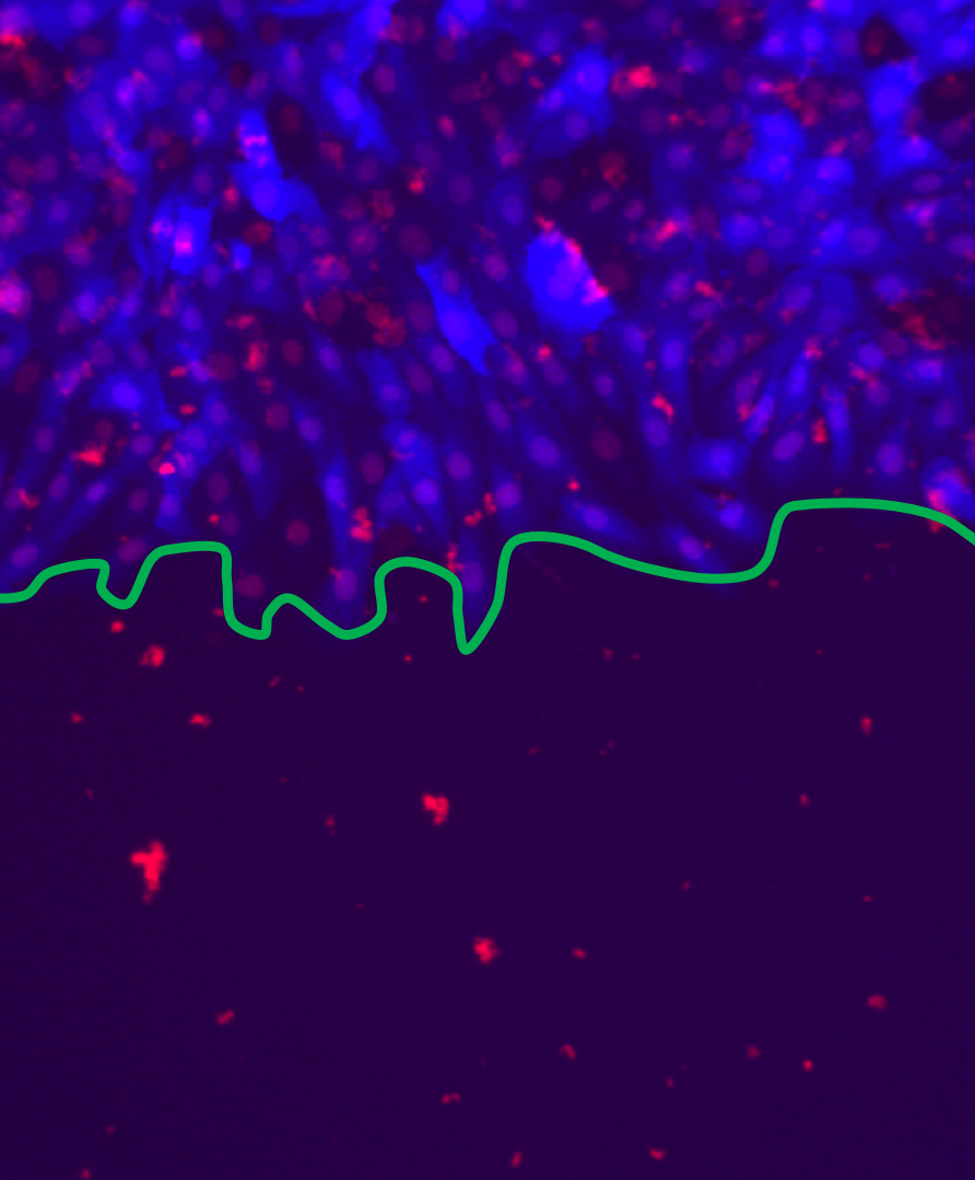
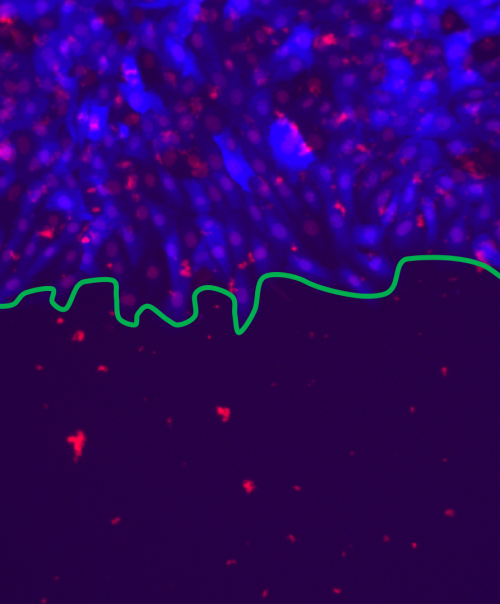
Microscopie à fluorescence d'un front (ligne verte) de cellules épithéliales de rat se propageant dans une boite de Pétri. Les cellules, avec le noyau (rouge) et le cytoplasme (bleu), forment un tissu bidimensionnel, servant à modéliser la cicatrisation.
--------
Fluorescent microscopy image of a proliferating cell front, whose edge is indicated in green. The rat epithelial cells, with cytoplasm (blue) and nuclei (red), form a 2-dimensional culture which can be used to model wound healing.


